Thoracic Spine Anatomy
Table of Contents

Mid Back
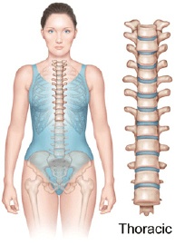
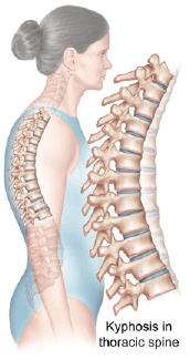
Thoracic spine is the central part of the spine, also called as dorsal spine, which runs from the base of the neck to the bottom of your rib cage. The thoracic spine provides flexibility that holds the body upright and protects the organs of the chest.
Spine is made up of 24 spinal bones, called as vertebra, of which, the thoracic region of the spine is made up of 12 vertebrae (T1-T12). The vertebrae are aligned on top of one another to form the spinal cord which gives the posture to our body. The different parts of the thoracic spine include bone and joints, nerves, connective tissues, muscles, and spinal segment.
Each vertebra is made up of round bone called as vertebral body. The protective bony ring attaches to each vertebral body and surrounds the spinal cord and forms the spinal canal. The bony ring is formed, when two pedicle bones join two lamina bones which connect to the back of the vertebral body directly. These lamina bones form the outer rim of the bony ring. When vertebrae arrange on top of the other, the bony ring forms a hollow tube which surrounds the spinal cord and nerves and provides protection to the nervous tissue.
A bony knob- like structure projects out at a point, where the two lamina bones join at the back of the spine; these projections are called as ‘spinous processes’ and the projections at the side of the bony ring are called as ‘transverse processes’.
Between each vertebra, there are small bony knobs at the back of the spine that connect the two vertebrae together, called as ‘facet joints’. Between each pair of vertebra, two facet joints are present, one on either side of the spine. The alignment of the two facet joints allows the back and forth movement of the spine. The facet joints are covered by a soft tissue called the ‘articular cartilage’, which allows the smooth movement of the bones.
On each side, the left and right side of the vertebra, is a small tunnel called ‘neural foramen’. The two nerves that leave each vertebra pass through this neural foramen. These spinal nerves group together to form a main nerve that passes to the organs and limbs. These nerves control the muscles and organs of the chest and abdomen. An ‘intervertebral disc’ is present in front of this opening which is made up of connective tissue. The discs of thoracic region are smaller compared to cervical and lumbar spine.
Connective tissue holds the cells of the body together and ligaments attach one bone to another. Anterior longitudinal ligament runs down to the vertebral body and the posterior longitudinal ligament attaches on the back of the vertebral body. A long elastic band, called as ‘ligamentum flavum’, connects the lamina bones.
The spine muscles are arranged as layers, strap-shaped spine muscle, called as ‘erector spinae’ makes up the middle layer of the muscle. The deepest layer of muscles attaches along the back of the spine bones and connects to the vertebrae. These muscles connect one rib to the other.
Spinal segment includes two vertebrae separated by an intervertebral disc, nerves that leave the spinal column at each vertebra, and small facet joints of the spinal column.

Other Major Segments
Important Structures of the Spine
- Vertebrae
- Intervertebral Discs
- Facet Joints
- Neural Foraminae
- Spinal Cord
- Nerve Roots
- Para spinal Muscles
- Spinal Segments


